BREAST EXAMINATION

regular breast screening starting from a relatively young age is extremely important in the prevention of breast cancer
breast cancer is the most common malignant tumour in women and occurs in about 1 in 8 Greek women
pre-existing breast cancer. If there has been breast cancer in one breast, it is more likely to develop new cancer in the other breast or in the treated breast
family history. Women who have a first-degree relative with breast or ovarian cancer or both are almost twice as likely to develop breast cancer
genetic predisposition. Mutations in genes, especially BRCA1 or BRCA2, lead to a higher risk of developing breast and ovarian cancer
excess weight. The higher is the BMI of women, the higher is the risk of breast cancer
hormones. Long-term exposure of a woman’s body to ovarian hormones affects her risk of developing breast cancer. Taking hormone replacement therapy after menopause increases the risk of developing breast cancer
social habits. Alcohol overconsumption can increase the risk of breast cancer.
race. White women are more likely to develop breast cancer than other races.
a diet rich in fruits and vegetables and low in saturated fats
vitamin A. It plays an important protective role in breast cancer
regular physical exercise. Moderate physical activity, even when started later, reduces the overall risk of breast cancer by at least 10%
breast screening is determined according to the age, individual and family history of each woman.
including:
breast self- examination
clinical examination of the breasts
breast ultrasound
mammography
it starts at a young age
it should be repeated every month between 7th and 12th day of the cycle
after menopause, you should choose a specific date and repeat the test every month on the same day
a woman herself can more quickly and easily recognize any change she sees, feels or feels in her breast
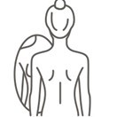 | Stand in front of the mirror with your hands down and observe your breasts. Then turn around and observe them on the side Pay attention to any changes in shape, size, obvious external lesions, presence of a lump, nipple inversion, skin lesions |
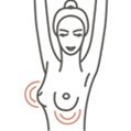 | Repeat the same checks with hands raised |
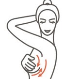 | Use your whole palm or even both fingers (index and middle) and feel your breasts in circular motions starting from the outside towards the nipple. Apply controlled pressure with circular movements and check for lumps. In the same way check the underarm area, with the hand lowered |
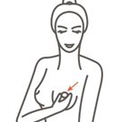 | Examine the nipple carefully Observe for visual alterations Check for fluid outflow before and after nipple pressure |
 | Lie down and place one hand behind your head Repeat the palpation in the same way Don’t forget the armpit check |
| Repeat the exact same checks on the other breast. |
you will sit on the examination seat and raise your arms to reveal any differences in the size and shape of the breasts and find any protruding masses
this is followed by palpation, where both hands are used to check each breast separately and the armpit
finally, pressure is applied to the nipple of the breasts to check for discharge
examination during which the breast is irradiated
during mammography, a minimal amount of radiation is released and repeating it annually does not increase any risk to the patient
if suspicious foci are identified on mammography then further imaging and invasive testing should be performed for a final diagnosis
digital mammography differs from traditional mammography only in the way the image is stored as a file on the computer, so with the use of appropriate programs these files can be enlarged by increasing their accuracy
age (years) | clinical guidelines for breast examination |
20-30 | annual clinical examination/self-examination shortly after the end of the period |
30-40 | annual clinical examination with ultrasound |
35-40 | digital mammography for future reference |
40-50 | digital mammography every two years with breast ultrasound and clinical examination |
50-75 | annual digital mammography with breast ultrasound and clinical examination |
>75 years | screening every two years, preferably with mammography and breast ultrasound |

